The Lamprobe is a multi-dynamic instrument for non-invasive and minimally-invasive, bloodless surgical removal of skin lesions.
Epidermal lesions, within the scope of practice of the Somatologist, can be safely and effectively removed on all skin types with selected probes.
Surgeons, Dermatologists and Doctors specialising in aesthetic medicine ensure extremely effective, minimally-invasive management of superficial to deep dermal lesions, with minimal co-lateral tissue damage and speedy tissue recovery, using the Lamprobe.
The future of skin care is moving away from invasive, burning devices that scar or pigment the skin. Clients and patients expect optimal results in era of advanced technology. The Lamprobe delivers results every time.
Based on radio wave and high frequency technology, the Lamprobe works on the principal of liquids being attracted to heat, released by various sized and shaped probes for the treatment of all types of different lesions. What distinguishes the Lamprobe from other devices, is the delicate yet sophisticated regulated intensity focusing on the lesion alone and not the underlying or surrounding skin.
The variety of probes are used to address all epidermal and dermal lesions.
Current intensity is specifically selected according to:
- Type of lesion
- Size of the lesion
- The site of the lesion within the epidermis or dermis
Treatment protocol is different for epidermal and dermal lesions.
The Lamprobe uses advanced technology for:
- Vaporising epidermal lesions similar to a microwave current
- Liquifying hardened lipids that have formed beneath the skin enabling effective removal
- Drying out sebaceous or fatty lesions
- Thermo-coagulating vascular or blood lesions
There are additional benefits:
- The removal of unwanted hair, irrespective of colour – Click here for more info on electrolysis with The Lamprobe
- The reduction of lines and wrinkles using radio frequency in collagen induction therapy – Click here for more info on CIT with The Lamprobe
- The treatment and management of acne and problematic skin
One device delivers it all and completes any aesthetic practice.
Benefits of Using Lamprobe
Treating skin lesions, blemishes and irregularities on skin has never been easier. The Lamprobe device allows for painless and efficient removal a variety of skin lesions, blemishes and irregularities. This non-invasive treatment offers immediate client satisfaction, with no blistering or down-time.
IMMEDIATE RESULTS, PRECISE TREATMENT, RAPID TREATMENT TIME, NO ANESTHETIC REQUIRED, NON-INVASIVE, SIMPLE AND EASY TO USE, TRAINING PROVIDED, VARIOUS PROBE SIZES, QUICK. SAFE. EFFECTIVE.
The future of Skincare
Skin Lesions
Remove all skin lesions with the Lamprobe and create flawless skin!
Everyone wants flawless skin. The Lamprobe is used to remove unsightly and uncomfortable skin tags, skin warts, broken capillaries, milia, acne pimples and DPN – instantly transforming the skin making the difference to all skin types. From light skin to Mediterranean, Asian, Indian and African – All skin irregularities can be cosmetically removed by skin therapists simply, quickly and effectively. Advanced lesions can only be removed by doctors.
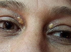
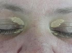
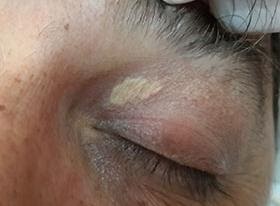
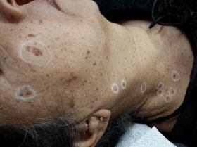
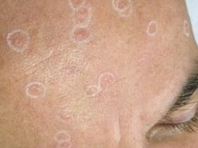
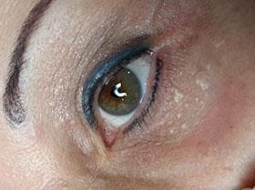
Do you have unsightly skin irregularities similar to these types of lesions?
Acne Pimples, Actinic/Solar Keratosis, Angiokeratomas, Basal Cell Carcinoma (Dr.), Blind Pimples, Broken Capillaries, Cherry Angiomas, Cholesterol Deposits (Large), Cholesterol Deposits (Small), Clogged Pores, Cutaneous Horn, Cysts, Dermatosis Papulosa Nigra / DPN, Dialated Capillaries, Fibromas, Milia, Moles (Dr.), Molluscum, Contagiosum, Neurofibromas, Pyogenic Granuloma, Raised Angiomas, Sebaceous Hyperplasia, Seborrhoeic Keratosis, Senile Warts, Skin Tag (With Stalk), Skin Tags (No Stalk), Solar Keratosis / Actinic Keratosis, Solar Lentigo, Spider Naevi, Squamous Cell Carcinoma (Dr.), Steatocytoma Multiplex, Syringomas, Telangiectasia, Warts, Xanthelasma
Remove these Skin Lesions with the Lamprobe.


Actinic /Solar Keratosis
These are the most common sun-related pre-cancerous growths noted in fair-skinned individuals. They are benign (non-malignant) and appear as crusty, dry scaly bumps that are rough textured like sand-paper. If left untreated, Actinic Keratosis can develop into Squamous Cell Carcinoma.


Xanthelasma
These are soft yellowish plaques of lipids usually found in the periorbital (around eyes) area. They are very tiny to medium in size and some may be raised. Some individuals are predisposed towards them. They are believed to be related to abnormal lipid metabolism.


Acne/ Blind Pimples/Congestion/Clogged Pores
Acne lesions are caused when the sebaceous glands located near the hair follicle become overactive, inflamed, infected with bacteria and fills up with pus.


Seborrhoeic Keratosis
These are non-cancerous benign skin growths that originate in the keratinocytes of the epidermis. These “barnacles of old age” vary in colour from light to black, are round or oval, slightly elevated and vary in sizes.


Dermatosis Papulosa Nigra (DPN)
These are fibromas that are prevalent in Fitzpatrick Skin Types V and VI (Indian, Asian and African skin tones). These benign lesions are darkly pigmented without scales, and are found most commonly on the face around the eyes, on the cheeks and the neck area. They can appear in adolescence and they increase with age.

Cholesterol Deposits (Large)
These are soft yellowish deposits of lipids usually found in the periorbital (around eyes) area. They are believed to be related to abnormal lipid metabolism.

Raised Angioma
These cherry red to purple coloured papules are also known as Campbell De Morgan spots or Senile Angiomas. Clusters of capillaries under the surface of the skin form a flat or raised round dome.


Telangiectasia / Broken Capillaries
Tiny superficial dilated blood vessels appear as red wavy lines just below the surfaces of the skin, mostly on the nose, cheeks and decollete area. Constant blood stimulation causes the thin elastic walls of the capillaries to vasodialate causing breakage. Trauma, lifestyle choices, those who suffer from allergies tend to be prone to Telangiestasia.

Spider Naevus
This is a common benign vascular lesion present in healthy adults and young children. It is characterized by a central red arteriole, or punctum, representing the body of a spider, with radical patterns of thin walled capillaries that look like wavy legs.


Sebaceous Hyperplasia
Chronic sun damage contributes towards these lesions resulting in enlarged sebaceous glands. They appear as a soft, yellowish papule with a cauliflower-like or doughnut-shaped appearance ranging in size from 2 to 3mm. They are usually solitary and appear on the forehead and cheeks, particularly on oily and asphyxiated skins.


Skin Tags
These are small fibromas that appear either single or in multiple formulation and are sometimes referred to as Achrochordon. They are commonly found on the neck, breasts and axillae in skinfolds and on eyelids. They can become inflamed with constant friction from necklaces or clothing.

Skin Tags With Stalk
Although unattractive, these are harmless growths that appear mostly on middle aged men and women. Skin tags are formed when the area of the outer layer of skin begins to overgrow and envelope collagen fibres, protruding from the surrounding skin. They vary in colour and shape and can be irregular.


Cholesterol Deposits (Small)
Tiny cholesterol deposits of lipids are believed to be related to poor lipid metabolism. They are very prominent under the eye and on the nose.

Milia
These plugs of sebum are covered with layers of Stratum Cornified Cells and are commonly found on the facial area where there is poor exfoliation for example on very oily or very dry skins. They can also occur during the healing of traumatic scars. Milia are commonly found on the forehead, cheeks and around the eyes.

Keratoses
These are non-cancerous benign skin growths that originate in the keratinocytes of the epidermis. These “barnacles of old age” vary in colour from light to black, are round or oval or irregular in shape, slightly elevated and vary in size. These are commonly called skin warts, however they do not have a virus present to validate the name.